Let me introduce you a particular species - Ascaris Lumbricoides. They are amazing, because the only two things they can do in life are - to eat and to reproduce. Why, you may wonder, this is amazing? Because they don't need anything else for life at all.


 Let's continue with dissection. The inner body organs are as well as simple as a particleboard. They are, as well, only responsible for digestion and reproduction.
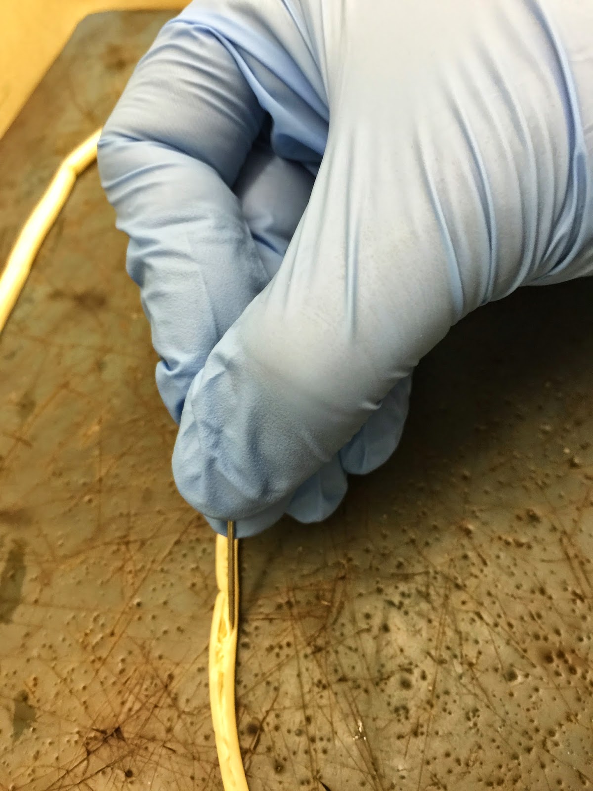
I started with a boy. To cut through the inner organs, I first had to pin the tail of a worm and very carefully cut his skin along the body. The process was pretty long and time consuming because I didn't want to screw up the organs.
For reference, Ascaris Lumbricoides is the giant roundworm of humans, growing to a length up to 35 cm. And yes, the probability that you have one is more than 75%.

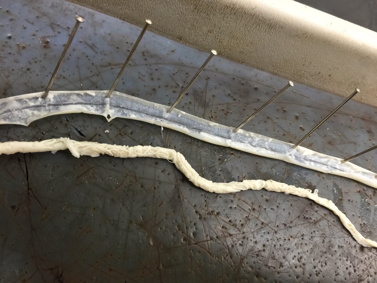
 After I cut from his head to his tail, I took part of the skin out so the organs could be seen. On the photo on the right, you can clearly see them. ==>
<== On the left image, I am showing the digestive tract, which is a tiny white line on the bottom of the body. All the other organs are responsible for reproduction.
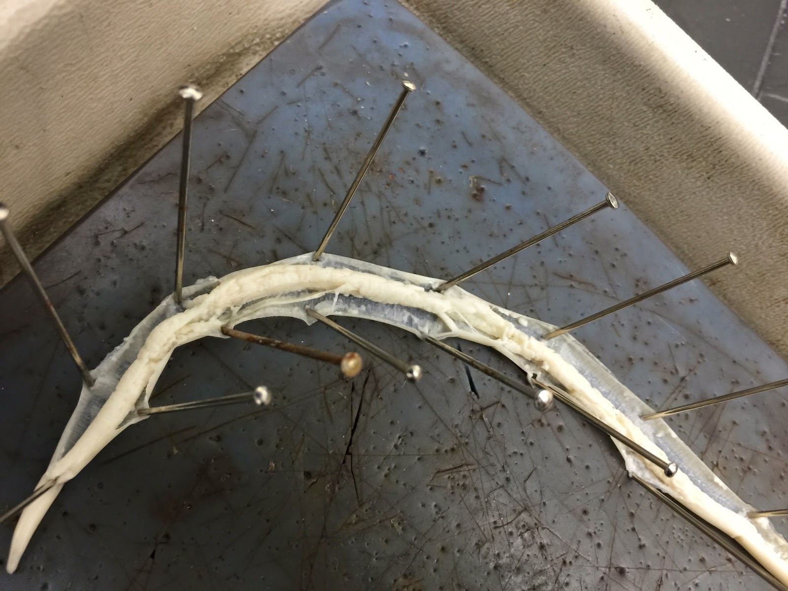
 For a better illustration, I basically skinned the boy so the inner organs could be better seen on the blue background of the workstation. ==>
Next, I continued with a girl, who was much easier to dissect because of the smaller size. The image below on the left illustrates a partially skinned female specimen, with inner organs being clearly visible. The image on the right shows her completely skinned.
 In a female specimen, it is much harder to identify special inner organs because of the body structure. At this point, I located a graph below (figure 9.16), showing each organ of the worms and explaining it. Try to compare the graph with photographs, and identify the organs.
Concluding at this point the topic of parasitic worms, I'll post the microscopic pictures essential for Biology laboratory:
 |
| Nector Americanus (adult) under |
 |
| magnification of x4 and x10 |
Nector Americanus
 |
| Trichinella, encrusted lavræ, in muscle (magnification of 4, 10 & 40) |


All three of trichinella spirals specimens illustrated above are as well parasites living in muscles.


Plantaria (the small white specimen), under magnification of x4 and x10.


Planaria injected (larger dark-green specimen) being shown under magnification of x10 and x4.


Chlonorchis sinesis, under magnification of x10 and x4.
Below, different parts of Tænia Pisiformis are shown under magnification of 4, as well as plain hydra.